

In-house developments and curated modules from external vendors — organized by modality. Integration runs uniformly through the platform.
From years of research with university hospitals: CNN-based methods deliver reproducible, quantitative measurements to support visual reading.
T2 / T1-CE lesions with inter-rater variability, compared to prior exams — compliant with the MAGNIMS-CMSC-NAIMS recommendations 2021.
Volumetry of the most important brain structures (whole brain, hippocampus, thalamus) on qualified scanners.
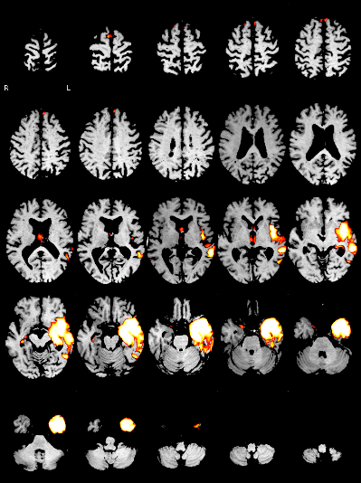
Voxel-based morphometry produces atrophy maps — for early assessment of focal atrophy and differentiation of neurodegenerative diseases.
The software is designed as a medical device for the computer-assisted detection of pulmonary nodules and fully meets the legal requirements for lung cancer screening.
and integration into the radiological workflow as key success factors.
The computer-assisted detection of pulmonary nodules on chest CT meets the legal requirements for lung cancer screening. With the introduction of early detection, AI support becomes mandatory.
Figures from internal validation against radiological readers. Please verify against current data before external use.
We develop our own algorithms continuously and integrate external AI per customer requirements — each with medical-device approval.
Lesions · volumetry · neurodegeneration · VBM
Hemorrhage exclusion
Nigrostriatal degeneration
Pulmonary nodules (screening-compliant)
ARIA · microbleeds (MRI) · RECIST
We evaluate external vendors against your requirements and advise you on the selection.
Request a consultation →